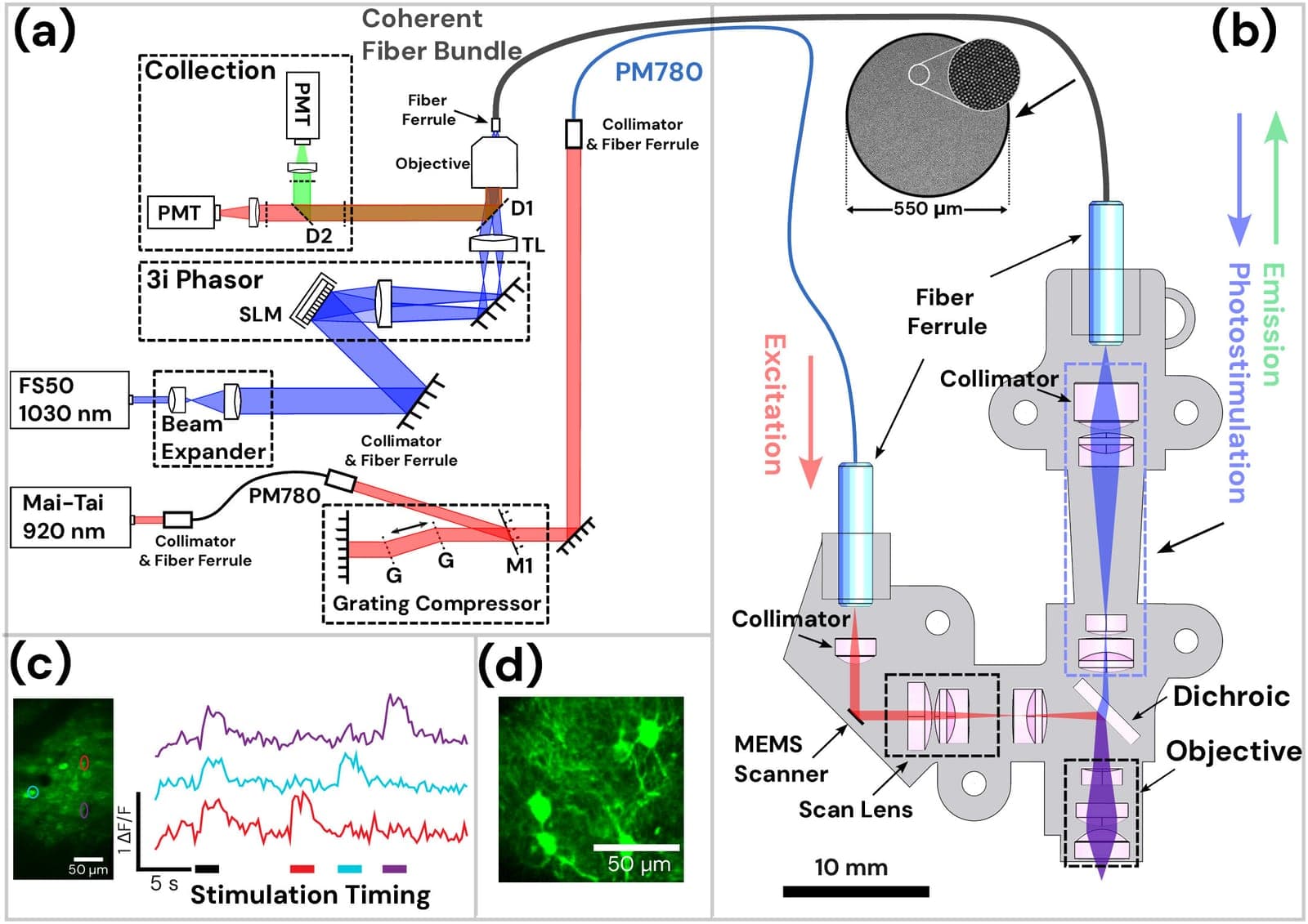
Researchers at the University of Colorado Anschutz and the University of Colorado Boulder have developed a miniature microscope that allows scientists to observe and activate individual brain cells during natural movement. The device, called Opto2P-FCM, combines high-resolution brain imaging with targeted light-based stimulation in a lightweight, head-mounted format that weighs 5 grams.
The new technology enables researchers to both monitor and manipulate the activity of specific neurons at the same time, according to the study published in the journal Optica. The device uses a novel dual-path optical design in which one pathway produces high-resolution images while a second delivers precisely patterned light that activates selected neurons without interfering with imaging. Separating the two functions allows each to be optimized independently, the researchers said.
Two-photon microscopy has long been considered the gold standard for producing detailed images of living brain tissue, but it typically requires subjects to remain stationary. While miniature microscopes have helped overcome some of those challenges, they have often sacrificed image quality or lacked the ability to precisely stimulate individual neurons. The new device overcomes these limitations, according to the study's senior author, Emily Gibson, Ph.D., associate professor of biomedical engineering at CU Anschutz.
The researchers said this technology could become an important tool for scientists studying disorders caused by disrupted brain circuits, including Alzheimer's disease, Parkinson's disease and epilepsy. By allowing researchers to observe and manipulate individual neurons in the same experiment, the system may help identify how healthy brain circuits function and how disease changes that. The current device is a prototype, but the authors are already working on future versions that will be smaller, lighter and faster while offering a larger field of view.