Optical imaging technique reveals immune cell metabolism in blood samples

Advanced optical imaging can detect the metabolic activity of individual immune cells in blood samples, providing information that traditional fluorescent labeling methods cannot reveal.

Oleh Sama News Agency
7 September 2026
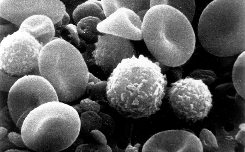
A black and white electron microscope image showing multiple round and irregularly shaped cells at high magnification, with smooth and textured surface details visible.
A scanning electron microscope image of immune cells reveals their varied morphology and surface structures, demonstrating the detailed cellular visualization that advanced optical imaging techniques can achieve in blood samples. (Phys.org)
1 menit membaca
Ukuran teks

A routine blood sample contains a diverse collection of immune cells that can provide clues about health and disease. Peripheral blood mononuclear cells, or PBMCs, are widely used in research on infections, autoimmune disorders, cancer and the immune system's response to treatment.

Scientists typically identify these cells using fluorescent labels attached to specific surface markers. However, those methods provide limited information about how the cells are functioning and can alter the cells during preparation.

A recent study reported in Biophotonics Discovery shows how advanced optical imaging can reveal previously inaccessible information: the metabolic activity of individual immune cells within a complex blood sample.


Optical imaging technique reveals immune cell metabolism in blood samples | Sama News Agency